


Humans and animals are attacked by these creatures, and infections are easily spread to each other through infected products, water and dirty hands.The emergence of in vivo parasites will help prevent prevention rules and careful adherence to personal hygiene, but even these measures do not guarantee 100% protection.The helmsman living in the human body can live in various organs, with different appearance, size and degree of harm to people.
Parasites in the human body
How many things are there when a person wanders around a doctor for years and can’t get rid of allergies, treats asthma, drinks sugar-down drugs and to no avail?Each of us has acquaintances like this who spend a lot of time treating various diseases and get no results.
Only in rare cases, when the doctor is very smart or very responsible, is he directing such patients to regularly analyzing feces and then…and then discovering parasites in the human body, which leads to dozens of diseases and no one fights to treat these pathology.
Contrary to established opinions, worms are not necessarily “prescription” in the intestines and can be detected by mediocre analysis of feces.Many parasites feel good in the lungs, heart, muscles, and even in the brain and eyes.
The famous malaria that once defeated returned again, the most dangerous parasitic disease that experts consider.Malaria parasites live only in the blood, and not every doctor can fully recognize the disease.
How parasites fall into human body
They fall into the body in different ways, often due to the use of infected water and food.
Grape eggs remain viable for up to 6 months and enter the body through toys, carpets, underwear and soft linen.Askarid eggs come into us through poorly washed vegetables and fruits.BBQ or homemade lard is a 95% guaranteed sternositis infection.
When bathing in freshwater reservoirs, bathing through air and dust, parasites penetrate our bodies with insect bites, which is the carrier of eggs.
Salt fish, sprout or caviar are the causes of worm infection, which reaches 12 meters in length and can live in the body under 25 years of age.Cases of infant infections become more frequent in parasites in the uterus.Dogs and cats can emit parasite eggs at distances of up to 5 meters through wet breathing.
You can not only get infected by your own hands, but also by dirty hands, but also by selling, chefs, waiters, parasites, travel by money and handrails.High concentrations of parasitic eggs are observed in products such as: bacon, smoked sausage, ham, sausage, any shape, beef, chicken, chicken, lamb, and even eggs, which are often infected by them.
Epidemiologists around the world are trying to fight this misfortune.For example, in the United States, every thousand pork cars are destroyed every thousand cars in order to verify Helminthias.This would develop with millions of dollars in losses, but otherwise it would not be possible.
There is no absolutely reliable way to disinfect meat, and the ordinary cooking process of larvae will not destroy.You can't keep your food purity by welding or frying meat, and a large number of parasite larvae can still penetrate your body.
Organs can live in parasites
The helm parasites are divided into two categories, corresponding to the activity locations in the donor:
- cramp: Worms that live in various gastrointestinal areas.There are about 100 species of intestinal parasites, and for each intestinal department, there are several species.The small intestine is ready to take Ascaris, Antrost, broadband and other less common "brothers".The small intestine will be with Pinworms, the dwarf chain and the rest of the "shared living space".Medical literature describes when a person is infected with several types of parasites simultaneously;
- Fabric:Worms are located in organs, tissues, and even in blood.Modern medicine successfully corresponds to parapathy (pulmonary), cyst disease (brain), echinococcosis (liver) and lymphocytosis (lymphocytes).Some worms pass through the body through the blood system and are randomly connected to any organ.If many eggs are introduced, it can infect the entire body.
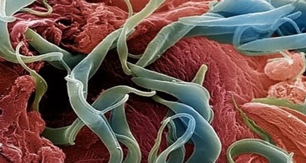
Snoes - How they look in the human body (photo of the worm)
Speaker- These are one of the most common human parasites, and are round worms (nematodes).Most commonly, pinworm infections occur in children, but are also found in adults.
The pins are white parasites, small and round.Individuals of females have dimensions: 8-13 mm in length, 0.5 mm in thickness, rectangular shape and straight tail pointing toward the end.
This feature of the female parasite tail explains its name - the word "cutting", "cutter".Men are much smaller: their length is 2-5 mm, their thickness is 0.2 mm, and their tail is curved, which is different from female pinworms.
The name of human invasion is intestinal taxation, which mainly occurs when it does not comply with personal hygiene rules (insufficient hand washing).Basically, PIN worms live in small portions of the intestine and upper part of the large intestine, but in some cases they can also migrate to other organs and organ systems.

The female worms fall into the human body orally and mate with the male representative of the nematode, migrating to the thick section of the intestine, where she gains the necessary lifespan and egg maturation from undigested food residues.
After 4 weeks, the female cutter began to migrate in the rectum at a rate of 12 cm per hour, crawled out of the anus, laying about 5,000-15,000 eggs under the hairy area, which after 4-6 hours was fully mature and ready for life cycle.
This process can be accompanied by itching, which encourages the infected person to comb the anus, thus helping further spread of parasites that fall from food under the nails into the hands of others (especially for children who are in close contact with each other, not always observing personal hygiene rules).
Pinworm infection from one person to another, through the dust of the parasite eggs, the patient touched it.Eggs can also be transferred to foods with cockroaches and flies.
Additionally, eggs are retained on linen, clothes, beds, which explains their rapid distribution.Since the worm's life cycle is very short, infections range from one person to one person, it is difficult to get rid of parasites because insulating from other nematode media is also necessary in addition to taking deworming drugs.
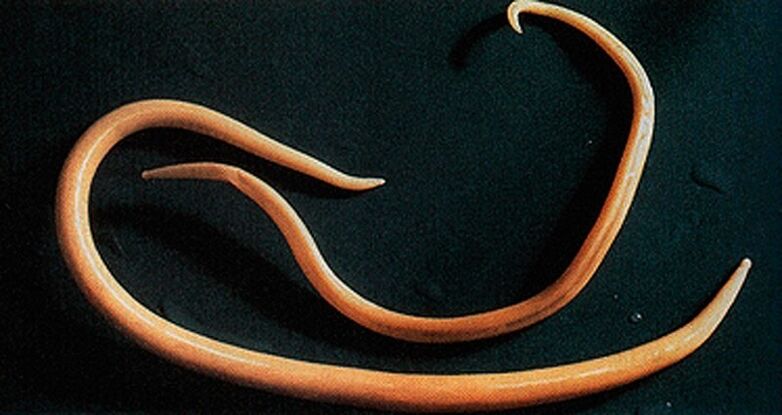
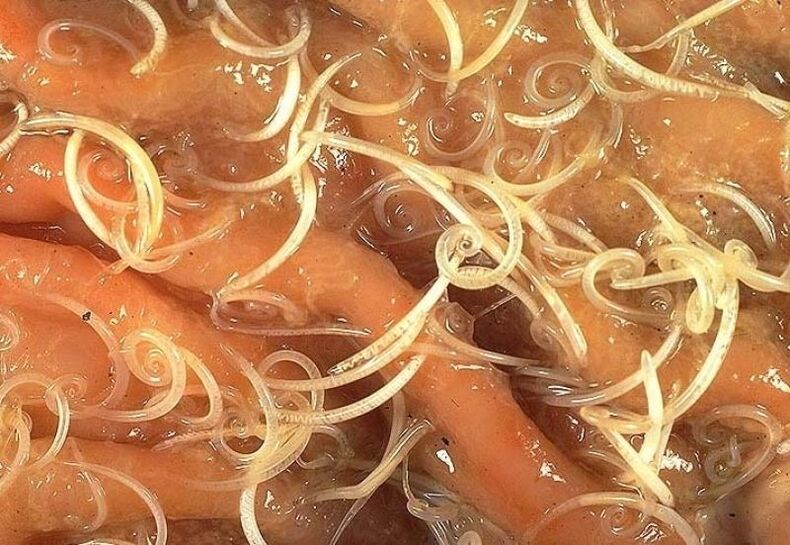
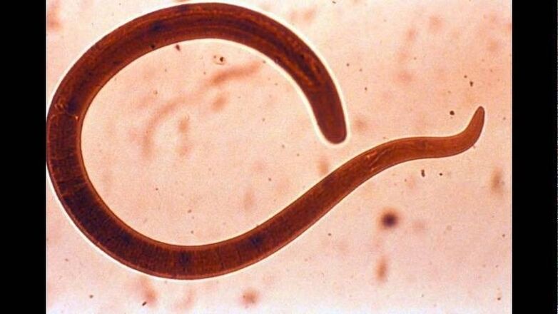
Askarides - How they look in the human body (photo of the worm)
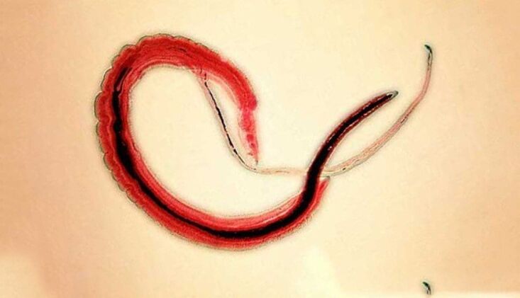
Ascarida- This is a large yellow color spindle-shaped large parasite, reaching 40 cm (female) and 15-25 cm (male) adult status.If there are no suction cups or other fixing equipment, Ascaride can move independently towards the food community.The eggs sent by female parasites are distinguished by feces.
Academic infections can occur in the presence of soil particles or unwashed vegetables and fruits.After the egg penetrates the intestines, mature larvae emit from it.
Then, on the intestinal wall, they reach the heart according to the blood flow, and then fall into the lungs from there.Through the pulmonary alveoli, the abdominal larvae through the respiratory tract penetrates into the oral cavity again.
In the intestinal phase it exists, assworms with spiral motility can penetrate even the narrowest pores.This characteristic of parasites often leads to the development of quite serious complications (mechanical jaundice or pancreatitis)
After repeated swallowing, the parasite reaches the small intestine, where it develops into an adult.The worm lived for 12 months, then died and stood out with feces.One or hundreds of people can live in the intestine of a master.
Allergens secreted by forrest granul gum can cause severe allergic reactions.A large number of adults can cause intestinal obstruction, and worms that penetrate the respiratory tract can sometimes cause suffocation.

Vlasov - Their appearance in the human body
BlacovyvThey often live in the southern region because the worm's eggs love heat.Most infections were observed in rural areas.Vlass - Head eggs live in the soil.
Invasion through hands, pollute the earth's particles, and poorly washed vegetables and fruits.Due to infection, disease occurs - three-point meningitis.Vrashv parasitizes in the intestine.This worm can cause anemia because it feeds on human blood and severe abdominal pain.
To diagnose the diagnosis of the hairy brain, the rectum and sigmoid colon are examined by a special device (venoscopy).Therefore, the accumulation of parasites in the intestine is found.The invasion treatment is long because the sore is protected by a thick shell.
Parasite eggs stand out in feces, but they are small and cannot always be seen even under a microscope.Eggs are only possible under very strong invasions.In shape, they look like buckets, with a brownish-yellow color.
On both sides, the egg is a hole.What do worms look like in feces?It is difficult to find them alive during bowel movements because the martyrs cannot live outside the human body for a long time.IV therapy can only be seen in the feces of white dead insects.
Liver Bacon - What it looks like in the human body
The parasite that causes myxamosis is a flat worm with a length of 7-20 mm.
In the acute stage of helminthosis, patients may experience soreness in the upper abdomen, elevated body temperature, nausea, muscle pain, diarrhea, diarrhea and rash.After the egg falls into fresh water (swallowed from the snail), the parasite larvae begin to develop.Then they penetrate the fish's body (carp, Crook carp, whales, cockroaches).
Human infection occurs when you eat infected fish that is not treated enough heat.The larvae of the liver bomb in the small intestine penetrated the bile duct and entered the gallbladder, and fixed there with two suction cups.
Symptoms of hepatitis, cholangioinflammatory, cholecystitis, impaired digestive tract, neurological diseases, weakness and fatigue show the chronic course of chronic myxopathy.The parasite is the driving force for the development of irreversible changes, and even after expulsion, the patient does not pass through chronic inflammatory processes and dysfunction.
Trichinella - Appearance in the human body (photo of the worm)
The pathogenic drug of Trichinellosis is a small round rudder with a length of 2-5 mm.When using fried meat (pork, bear cubs, wild boars), infection occurs.Within 3-4 days, parasite larvae penetrate into the intestine, maturing the state of sexually mature individuals.
The life expectancy of the worm is 40 days, after which the parasite dies.Drive the intestinal wall, the larvae penetrate the blood and carry it in all organs of the body, settle into the muscles.In this case, the bending of the respiratory and facial muscles, as well as the limbs, is usually affected.
In the first few days after the invasion, the patient complained about abdominal pain.
Then, after about 2 weeks, the body temperature rose to 39-40 s, the rash appeared on the skin, muscle pain developed, and the face was swollen.
During this period, in the case of large-scale infections, the risk of death is high.About a month later, recovery occurred.The parasite is encapsulated in a spiral form, after which it dies within two years.

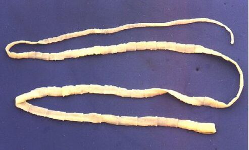
Wide tape - its appearance in the human body
This is one of the largest worms, with a length of 10-20 meters.The disease caused by this parasite is called dicytosis.The worm development cycle begins with freshwater fish or crustaceans.
Arriving at the small intestine, the parasite attaches to its walls and grows to a sexually mature individual on 20-25 days.
The larvae enters the human body, which is the ultimate owner of a wide ribbon with caviar or infected fillets.
Difillobotisos was performed in the context of digestive tract and B12-deficient anemia.
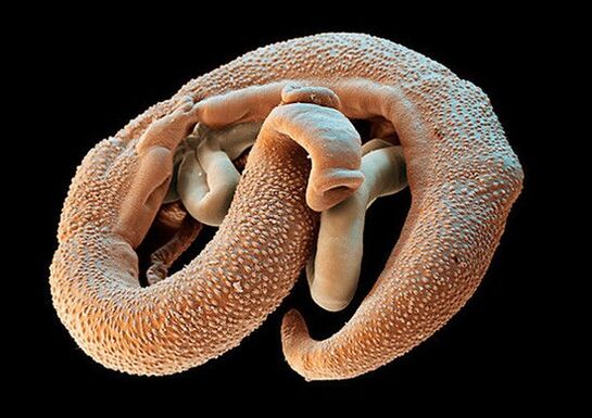
echinococcus-Its appearance in a person's body
For this parasite, one person is the middle host.Worms are parasitic in the human body in the form of Finns.The ultimate owner of echinococcus is a wolf, dog or cat.
The infection is in contact with animals and environmental objects in a digestive manner and carries a handful of echinococcus eggs.After entering the intestine, tumor balls (six-leaf larvae) develop from it.From the intestine, they penetrate the blood and carry it throughout the body.
The "favorite" places for worms parasitizing are the liver and light.Settled among these organs, the larvae became Finnish (Epithecus cyst), which gradually increased in size and began to destroy nearby fabrics.
Usually, echinococciosis during diagnosis is mistaken for benign or malignant tumors.In addition to mechanical exposure (squeezing organs and blood vessels), sometimes echinococcal cyst rupture occurs.This condition can cause toxic impacts or the formation of multiple new cysts.
Alveolar - How it looks in the human body
The parasite is considered to be a variety of echinococcals and is one of the most dangerous causes of helminthiasis (pneumococcal), similar to cirrhosis and liver cancer in severity.Infection occurs as intestinal bulbs (eggs with mature larvae) penetrate into the intestine.
This parasite is considered to be various sprigillus and is one of the causes of one of the most dangerous helminths (alveolar disease)
There, the embryo comes out of the egg and is introduced into the intestinal wall and penetrates into the blood.Furthermore, as the blood flows, the parasite spreads through all tissues and organs of the body (usually located in the liver).The main stage of development begins with larvae (multi-chamber bubbles, monsoon cysts)).
Each chamber has an embryonic head of a parasite that continues to develop gradually.Toilets are very radical formations that grow constantly due to the increase of air bubbles and are able to sprout to the liver like cancer metastasis.
Necrotic changes in tissue near tissue caused by impaired blood vessel operation.In the nearby structure opens, the alveoli form the content of multi-chamber bubbles.This can last for years, in case of mandatory surgical intervention.
Schistosoma - The appearance of the human body
Schistosoma -a blood-couple factor, belonging to the trematodes class, depending on the type that causes the various schistosomes.This is a separate worm with a length of 4-20 mm and a width of 0.25 mm.The body of the spirochete is equipped with a second suction cup - the mouth and abdomen, which are close to each other.The sketched women are longer and thinner than men.There is a longitudinal groove in the male body and he holds the female.Their eggs are 0.1 mm in diameter and are oval in shape, and on the surface of one of the rods is a large spike.
In the role of the final owner, the human worms of blood clots choose humans, in their organisms, they are parasitized in small veins of the colon, abdominal cavity, uterus, bladder.Worms feed on blood and partially absorb nutrients through their epidermis.Eggs with spots are transported to the intestines and bladder, where they mature and stand out in feces or urine.In freshwater waters, larvae-Miucidius, in which the intermediate host-mollust comes out of the egg.In the body of molluscs, the palmaria develops into a crack within 4-8 weeks.
Pork flavor - the appearance of the human body
Like a bull, the pork tape worm has 4 suction cups, but in addition to that, the worm's body is equipped with double hooks.The length of the Strogil is two to three meters.The ovary of the pork tapeworm has a three-mixed ovary, with 7 to 12 branches on both sides of the uterus.One characteristic of this worm is the ability of segments to crawl out of the anus.Once the outlet is exported to the external outlet, their shells dry and break, so the worm eggs enter the external environment.The middle master of the tapeworm can be a pig and a person.
The main owner is one person.Intestinal parasites in humans include pork tapeworms, which are located in the patient's intestines, where he lays eggs.When invasive meat is used, an infection occurs.
Which doctor is in contact with the worm
If you suspect an infection with a worm, it is necessary to consult a doctor, infectious disease expert, Helminthologgy Or parasitologist.You can contact any parasite or protozoa with an infectious disease specialist related to the infection.
You can contact a psychologist only if you accurately suspect infection through parasites (Pinworms, Ascarides, Contamination, Capillaries, Opisthorchiasis, etc.).
In case of suspected infection of protozoa, you can contact the parasitologists - Lamblia, Toxoplasmes and Amoebas.
Furthermore, if the parasite is not in the lumen of the intestine or stomach, but in other organs (e.g., lungs, liver), you can contact experts involved in the diagnosis and treatment of the disease in that organ.That is, for example, with mycoticosis, you can also contact a gastroenterologist or hepatologist and a pulmonary doctor.